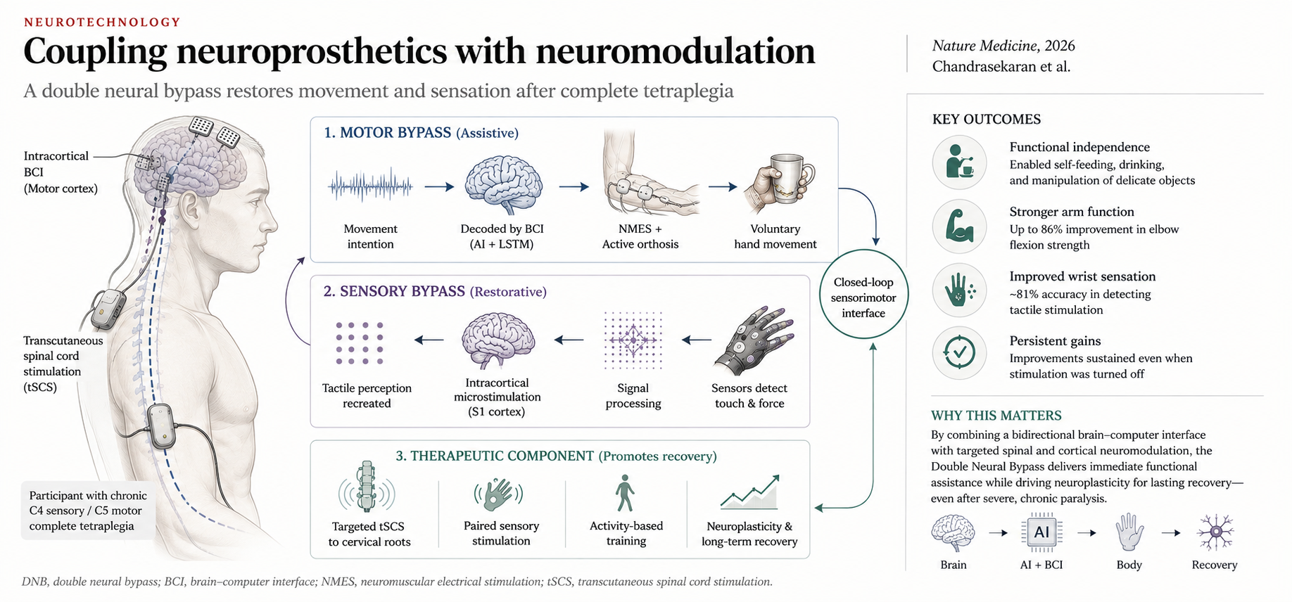
Researchers have successfully developed a “double neural bypass” (DNB) system that restores both immediate sensorimotor function and lasting neurological recovery in a patient with chronic, complete tetraplegia.
There is a quiet assumption baked into most neuroprosthetic research: the device is the therapy. Turn it on, function returns. Turn it off, it’s gone. The person becomes, in a very literal sense, dependent on the hardware for as long as they want the function back.
A paper just published in Nature Medicine — “A neuroprosthesis for restoring hand movement and sensation in a person with complete tetraplegia,” from Chandrasekaran, Wandelt and colleagues — quietly breaks that assumption. And it does it in the population where breaking it matters most: a person with a chronic, complete C4 sensory/C5 motor spinal cord injury, thirteen months out from a diving accident, with no voluntary hand movement and no tactile sensation below the level of injury. This is not the population where spontaneous recovery happens. It’s the population where “nothing more to do” has historically been the honest clinical answer.
The question nobody was quite asking
Brain-computer interfaces that let a paralyzed hand move again aren’t new — the same group’s earlier work, and Ajiboye and colleagues’ 2017 Lancet paper, established that decoding intention from motor cortex and driving muscle stimulation could restore functional grasp in complete tetraplegia. What was missing from that first generation was sensation — you can move an object you can’t feel — and, more fundamentally, any mechanism for the intervention to do something the nervous system would keep after the electrodes were switched off.
This paper’s real contribution isn’t that it moves a hand and restores touch simultaneously, though it does both. It’s that it treats “restore function now” and “promote lasting recovery” as two separate engineering problems requiring two separate solutions, built into the same device, running on two different timescales.
Two loops, two timescales
The system — the authors call it a double neural bypass, or DNB — has an assistive module and a therapeutic module, and the distinction is the whole argument.
The assistive loop reads from two microelectrode arrays in primary motor cortex, decodes movement intention using a recurrent neural network trained once and then locked — no retraining — and drives the participant’s own hand via neuromuscular stimulation and a custom 3D-printed orthosis. Grasp force, which turns out to be poorly regulated by cortical decoding alone because motor cortex activity is strongest at movement onset and offset rather than sustained through a hold, is stabilized instead by a nested deep reinforcement learning agent. Tactile information from force sensors in the hand is routed back as intracortical microstimulation in somatosensory cortex. The result: the participant can grasp a hollow eggshell without crushing it, drink from a cup, feed himself, and — in a moment that reads less like an engineering demo and more like the actual point of the paper — grasp and feel his sister’s hand with his own.
The therapeutic loop is where this paper distinguishes itself from everything before it. Targeted transcutaneous spinal cord stimulation, delivered to specific dorsal cervical roots identified by EMG recruitment mapping, was paired with activity-based training. Separately, a novel intervention the authors call “cortical mirroring” delivered patterned stimulation to somatosensory cortex designed to replicate the spatial signature of neural activity recorded during natural touch, paired with peripheral vibrotactile input.
Neither of these is meant to move the hand in real time. Both are meant to change the nervous system’s baseline.
The numbers that matter
Spinal stimulation alone, paired with activity-based training, produced a 61% and 25% increase in volitional elbow flexion force within fifteen weeks on the right and left sides respectively — climbing to 86% and 62% by roughly thirty-five weeks. That’s not a stimulation-on effect measured mid-session; that’s a standing gain in strength, measured off-stimulation, that let the participant bring both hands to his face for the first time since his injury.
Tellingly, the same intervention did nothing for the hand muscles or for tactile sensation. Spinal stimulation, it turns out, amplifies what residual substrate is already there — it doesn’t manufacture substrate that doesn’t exist. That negative result is arguably as important as the positive ones, and it’s the kind of finding that only shows up when you’re honest enough to test the intervention where you expect it to fail.
Sensation was the harder problem, and it needed the combined intervention. After thirty-nine weeks of spinal stimulation alone produced no change in wrist sensitivity, adding cortical mirroring did — the participant went from no sensation to reliably perceiving forces as low as ten grams on his previously insensate wrist, a gain that was still measurable more than two months after stimulation stopped. Electrode-level recordings back this up: the specific S1 electrodes involved in the mirroring intervention showed significantly increased responsiveness to real touch afterward, with that modulation continuing to build for over ninety minutes after stimulation had ended. Outside the lab, the participant reported being able to scratch his own face — and to feel his dog’s fur on the wrist that, a year earlier, felt nothing at all.
An old idea, borrowed and rebuilt
If this mechanism sounds familiar, it should. Pairing peripheral input with central stimulation to lower a threshold and induce a lasting change in cortical responsiveness is the same logic underlying paired associative stimulation and rTMS/tDCS priming protocols — just executed here with intracortical precision, and with the spinal cord doing double duty as an excitability gate for peripheral signals that the injury had otherwise attenuated. The authors are explicit that this is the mechanism they believe is at work: spinal stimulation raises interneuronal excitability, and cortical mirroring supplies a network-level template of what touch should look like, delivered while that window is open.
What’s genuinely new is the “network-level” part. Most biomimetic stimulation work has focused on temporally patterning one or a few electrodes to mimic natural firing at a single receptive field. Cortical mirroring instead recreates the broader spatial pattern of activity across the cortical population — including, plausibly, neurons adjacent to but not strictly part of the touch percept itself. Whether that broader net is doing the real work, or whether it could be made more efficient by combining it with the finer temporal patterning of biomimetic approaches, is an open question the authors raise themselves.
What this doesn’t prove
This is one participant. The authors are careful not to overclaim, and neither should we. We don’t know how this generalizes across lesion levels, injury chronicity, or the amount of residual substrate a given patient has — and the negative result with hand-muscle spinal stimulation is a pointed reminder that “more stimulation” doesn’t rescue a circuit with nothing left to amplify. The system also requires a specialized, multi-person team to operate; this is not, today, something that scales to a general SCI rehabilitation program. The mechanistic story — spinal gating plus cortical templating — is well-supported by the electrophysiology but remains inferential rather than directly confirmed.
Why this matters beyond spinal cord injury
The detail I keep returning to is a deliberate design choice buried in the discussion: the authors chose a transcutaneous, rather than epidural, approach to spinal stimulation specifically because it lowers the surgical burden and makes the therapeutic module more portable to other conditions — stroke among them. That’s the tell. This paper isn’t really about spinal cord injury. It’s a proof of concept for a more general idea: that neuromodulation aimed at inducing lasting change and neuromodulation aimed at restoring function in real time are not competing uses of the same real estate on the nervous system. They are two different interventions, operating on two different clocks, that belong in the same treatment architecture.
That is not a foreign idea to anyone working across Transcranial Magnetic Stimulation, tDCS, taVNS and neurofeedback in the same clinical program. It is, in fact, the working premise. What this paper offers is a rigorously instrumented, single-case demonstration of what it looks like when that premise is taken all the way to its logical conclusion — built into one device, tested against its own negative results, and left running long enough to show what remains when it’s switched off.
The eggshell is the demonstration. The dog’s fur is the discovery. The field’s task now is to figure out how much of the second effect can be engineered on purpose, in more people, with less hardware.
Chandrasekaran, S., Wandelt, S.K., Jangam, A. et al. A neuroprosthesis for restoring hand movement and sensation in a person with complete tetraplegia. Nat Med 32, 2591–2601 (2026). https://doi.org/10.1038/s41591-026-04498-0

Leave a Reply